 KSOEM
KSOEM

Articles
- Page Path
- HOME > Ann Occup Environ Med > Volume 38; 2026 > Article
- Original Article Evaluating the efficacy of prednisolone for silicosis with progressive massive fibrosis in Australia: a pre-post quasi-experimental study
-
Hayley Barnes1,2,3,4,*
 , David P. Nadebaum5,6, Daniel Niewodowski1, J. K. Khoo1, Yuan Z. Lim7, Bradley J. Gardiner3,8, Tiffany Lin1, Martin H. Cherk5,9, Miranda Siemienowicz3,10,11, Jyotika D. Prasad1,3, Ryan Hoy1,2
 -
Annals of Occupational and Environmental Medicine 2026;38:e16.
DOI: https://doi.org/10.35371/aoem.2026.38.e16
Published online: June 4, 2026
1Department of Respiratory Medicine, Alfred Health, Melbourne, Australia
2Monash Centre for Occupational and Environmental Health, Monash University, Melbourne, Australia
3School of Translational Medicine, Monash University, Melbourne, Australia
4Department of Medicine, University of California, San Francisco, CA, USA
5Department of Nuclear Medicine & PET, Alfred Health, Melbourne, Australia
6Department of Neuroscience, Monash University, Melbourne, Australia
7Department of Rheumatology, Alfred Health, Melbourne, Australia
8Department of Infectious Diseases, Alfred Health, Melbourne, Australia
9Faculty of Medicine, Monash University, Melbourne, Australia
10Department of Radiology, Alfred Health, Melbourne, Australia
11Northern Imaging Victoria, Northern Health, Epping, Australia
- *Corresponding author: Hayley Barnes Monash Centre for Occupational and Environmental Health, 553 St Kilda Rd, Melbourne, VIC, 3004, Australia E-mail: Hayley.Barnes@monash.edu
• Received: February 20, 2026 • Revised: May 17, 2026 • Accepted: May 27, 2026
© 2026 Korean Society of Occupational & Environmental Medicine
This is an Open Access article distributed under the terms of the Creative Commons Attribution Non-Commercial License (https://creativecommons.org/licenses/by-nc/4.0/) which permits unrestricted non-commercial use, distribution, and reproduction in any medium, provided the original work is properly cited.
- 1,314 Views
- 54 Download


Abstract
-
Background There has been a resurgence of silicosis, particularly related to artificial stone. There are currently no treatments for silicosis beyond lung transplantation for end-stage disease. This study aimed to evaluate the efficacy of prednisolone in people with artificial stone-associated silicosis–progressive massive fibrosis (PMF).
-
Methods This was a pre-post quasi-experimental study, assessing 3 months of prednisolone in adults with artificial stone–associated silicosis with PMF. Outcomes were assessed at 3 and 12 months.
-
Results Seven participants completed the study. Baseline positron emission tomography (PET) scans demonstrated increased fluorine-18 fluorodeoxyglucose (18F-FDG) uptake in areas of PMF in all participants. All participants had a significant reduction in maximum standardized uptake value (SUVmax) following prednisolone at 3 months (pre-treatment mean SUVmax 6.7 [standard deviation (SD): 2.6], post-treatment mean SUVmax 4.2 [SD: 1.0], p = 0.012). There was also a non-significant reduction in the % of total lung parenchyma with SUV >1 (49.7% [SD: 36.4] to 45.8% [SD: 28.4], p = 0.527) and a significant reduction in SUV >2.5 (7.0% [SD: 6.3] to 2.4% [SD: 2.2], p = 0.038). There was a non-significant reduction in computed tomography ICOERD well-defined opacity profusion scores and large opacities. There was no significant difference in lung function or St. George's Respiratory Questionnaire. There were no serious adverse events.
-
Conclusions There are high levels of inflammation in silicosis-PMF as evidenced by 18F-FDG PET. Short-term prednisolone reduced 18F-FDG PET activity. This suggests a possible therapeutic pathway for people with silicosis-associated PMF in a population with no current treatments. Further research is required to determine the most appropriate immunosuppressive strategy and further assess longer-term outcomes.
BACKGROUND
Silicosis is an interstitial lung disease (ILD) caused by the inhalation of respirable crystalline silica (RCS) particles. In recent years there has been a rapid increase in cases attributable to the fabrication of artificial (engineered) stone benchtops in Australia,1 Spain,2 Israel,3 and United States.4 Patients with silicosis may be asymptomatic, or develop breathlessness, cough, chest pain, reduced exercise tolerance, and respiratory failure, with worse symptoms more likely in those with progressive massive fibrosis, and a life expectancy as short as seven years.5
Silicosis results from direct cytotoxic effects of RCS and stimulation of inflammatory and fibrotic pathways leading to parenchymal damage and a varied spectrum of disease. Inhaled crystalline silica particles reach the distal bronchioles and alveoli of the lung, where they are engulfed by resident alveolar macrophages and endocytosed by epithelial cells lining the airways. This leads to chronic activation of the nucleotide-binding domain, leucine-rich–containing family, pyrin domain–containing-3 (NLRP3) inflammasome pathway, resulting in maturation and release of multiple downstream effector cells and cytokines, including interleukin (IL)-1β, and IL-18, Th1 lymphocytes, eosinophils, mast cells, and fibroblasts.6 In an attempt at repair, transforming growth factor β (TGF-β) and IL-6 stimulate differentiation of Th17 lymphocytes, which promote granuloma formation via the production of IL-17.7 Granulomas and inflammatory cells aggregate to form silicotic nodules. Permanent lung fibrosis and airway remodeling is driven by the pro-fibrotic mediator TGF-β, a key effector of collagen deposition and turnover in fibrotic lung diseases (Fig. 1). In addition to silica, pigments and resins contained within artificial stone are respiratory irritants that likely contribute to inflammation and oxidative stress.8
Management of silicosis requires avoidance of further exposure to RCS dust. Even following exposure cessation, many patients will exhibit progressive disease. Despite its increasing prevalence, there are no routinely recommended treatments in clinical practice beyond lung transplantation for end-stage disease.9
Recent novel imaging studies demonstrate high levels of inflammation in regions of progressive massive fibrosis (PMF) associated with artificial stone silicosis.10 Fluorine-18 fluorodeoxyglucose (18F-FDG) positron emission tomography (PET)/computed tomography (CT) demonstrates significant uptake in areas of PMF, which challenges the notion that silicosis-PMF is completely fibrotic and irreversible, and suggests possible therapeutic targets. In vitro and murine studies have demonstrated attenuated inflammation and fibrosis with immunosuppressive therapy,11 but clinical trials in humans are lacking.
Prednisolone is a commonly used oral corticosteroid that has a broad range of immunosuppressant effects. These include down-regulation of cytokines (including IL-1β, tumor necrosis factor α, IL-6, -8, -12, and -18),12 reduction of activation via the mitogen-activated protein kinase/ERK and nuclear factor-кB pathway,13 improve phagocytosis, and reduce oxidative stress.14 Prednisolone has been utilised in a multitude of conditions, including as first-line therapy for sarcoidosis, asthma, and autoimmune diseases.15 As a short-term therapy, it has few side effects, is orally available, low-cost, easy to access, and its use is familiar to many around the world.
Therefore, we conceived the STOP study (Silicosis Treatment of Prednisolone), of which the objectives were to assess the safety, feasibility, and efficacy of prednisolone in people with artificial stone-associated silicosis.
METHODS
This prospective quasi-experimental study assessed outcomes before and after the intervention. Participants were recruited from the Alfred Occupational Respiratory Clinic, Alfred Hospital, Melbourne, Australia (from 07/2023 to 10/2023), which screens workers from the stone benchtop industry and manages those who develop silicosis due to this exposure.
Silicosis was determined using a multidisciplinary process incorporating exposure history, CT appearance as assessed by specialist Thoracic Radiologists, and endobronchial ultrasound (EBUS) lymph node sampling or transbronchial biopsy. Participants were invited if they had complicated silicosis with PMF (PMF - as defined by one or more large opacity ≥10 mm in cross-sectional diameter) due to artificial stone dust exposure. Participants were excluded if they had ongoing exposure to silica, uncontrolled diabetes mellitus, untreated infections, including tuberculosis, any history of serious infections, on immunosuppressants for any other condition, and those with asthma. Sample size was based on the feasibility of recruitment. Those from culturally and linguistically diverse backgrounds were actively recruited.
After obtaining consent, participants were administered 25 mg oral prednisolone daily for 3 months, followed by a short tapering course over two weeks. To reduce the risk of Pneumocystis jirovecii pneumonia (PJP) infection, participants were also prescribed PJP prophylaxis (trimethoprim 160 mg/sulfamethoxazole 800 mg three times a week) for 3 months, and esomeprazole 20mg daily for 3 months to reduce the risk of reflux and gastrointestinal bleeding. Participants were counselled on possible adverse events and provided written information.
As the main objective of this study was to assess the effect of immunosuppression on inflammation in silicosis-PMF, the primary outcome measure was the change in maximum standardized uptake value (SUVmax) on 18F-FDG PET. Additional PET parameters evaluated included total lung glycolysis (TLG), % lung >1 SUV (1 SUV considered normal parenchyma16), and %lung >2.5 SUV (2.5 SUV average uptake in idiopathic pulmonary fibrosis [IPF]17).
Participants were asked to fast and refrain from vigorous activity for at least 6 hours before imaging. Administered 18F-FDG activity was 3 MBq/kg up to a maximum of 300 MBq. Pre and post prednisolone PET/CT scanning was performed on a Discovery 710 PET/CT scanner (GE Healthcare, Milwaukee, WI, USA) with respiratory gating and a scan range from upper neck to upper abdomen, 60–75 minutes after injection of 18F-FDG. Low-dose co-registered CT was used for anatomical localization and attenuation correction. 12-Month follow-up scans using the same protocol were performed on a Vision 600 PET/CT scanner (Siemens Healthcare, Knoxville, TN, USA) due to interval camera replacement. All images were reviewed independently by two nuclear medicine specialists experienced in 18F-FDG PET/CT (DM and MC), blinded to all other investigation results.
Quantitative analysis of PET/CT images was performed using MIM Software (Cleveland, OH, USA). Lung segmentation was performed using the low-dose CT images and the region grow function (left and right lungs delineated with their respective tools). The 3D brush function was used for manual correction if required. This included the removal of the trachea/main bronchi and any areas of 18F-FDG flaring from the left ventricle or hilar/mediastinal nodes into adjacent lung, and conversely the inclusion of any areas of PMF not captured by automatic total lung segmentation.
Quantitative PET parameters were derived from the total lung region-of-interest, which included areas of progressive massive fibrosis. Measurements included total lung SUVmean, SUVmax, TLG, and the percentage of total lung volume with SUV above the pre-defined thresholds of 1 and 2.5.
– Change in lung function: (including forced expiratory volume in 1 second [FEV1], forced vital capacity [FVC], and diffusion capacity for carbon monoxide [DLCO]), using standardised criteria.18
– Change in inflammatory/autoimmune markers (angiotensin-converting enzyme [ACE] level, erythrocyte sedimentation rate)
– Change in health-related quality of life measures: St. George’s Respiratory Questionnaire (SGRQ). The SGRQ is a 50-item questionnaire designed to measure the impact on overall health, daily life, and perceived well-being. Scores range from 0 to 100, with higher scores indicating more limitations. The SGRQ has been described in silicosis, and scores are associated with the degree of lung function impairment.19,20
– Change in CT ICOERD (International Classification of High-resolution Computed Tomography for Occupational and Environmental Respiratory Diseases) scores. For this study, paired CT scans were scored using a modified ICOERD criteria. ICOERD uses a 4-point grade scale (0–3) to quantify the extent (profusion) of the following parenchymal abnormalities: well-defined rounded opacities (RO), irregular and/or linear opacities (IR), emphysema, ground glass opacities, honeycombing, and large opacities (LO). A grade is assigned for each of the upper, middle, and lower zones of both lungs. The ICOERD criteria are designed for high-resolution routine dose CT. The CT used in FDG-PET for co-registration has significant technical differences, including significantly lower resolution and more frequent obscuration of lung parenchyma by physiological atelectasis. As such, the presence or quantity of ground glass opacity or ‘P’ category of rounded opacities (<1.5 mm) could not be assessed. It should be noted that accelerated silicosis relating to artificial stone frequently manifests as centrilobular pure ground glass nodules, and this finding is not appreciable on this modality. Intralobular reticulation was scored, but sensitivity for this finding is expected to be much lower than on diagnostic CT for the technical reasons described. The summed score for this study includes the domains: RO, IR, and LO.
Outcomes were measured prior to and following 3 months of prednisolone. A repeat PET scan and lung function tests were performed 12 months following cessation of treatment. Adverse events were also assessed. Occupational and exposure histories were collected using a previously described exposure assessment instrument.21
Continuous variables (e.g., FEV1 % predicted, FVC % predicted, and DLCO % predicted) were analyzed using a paired t-test (dependent t-test) where data were parametric or a Wilcoxon signed-rank test where data were non-parametric. Categorical data were analyzed using McNemar’s test. The mean average change scores are presented. Correlation was calculated using Pearson correlation. Where data were missing, we did not impute data as we could not be certain that this was not random and this was a small sample size. Analyses were performed using SPSS version 30 (IBM Corp., Armonk, NY, USA). This study was conducted and reported in accordance with TREND (Transparent Reporting of Evaluations with Nonrandomized Designs) guidelines.22
The study protocol was reviewed and approved by the Alfred Ethics Board (187/23). Informed consent was submitted by all subjects before they enrolled in the study. The trial was prospectively registered (ACTRN12623000622606).
RESULTS
Eleven participants were screened, and seven participants completed the study. Two participants were excluded because they had a history of tuberculosis and current infection could not be excluded (declined bronchoscopy), and two participants completed the screening process but withdrew prior to the commencement of the intervention. Baseline demographics of included participants are presented in Table 1. Once enrolled, all participants completed the study.
Pre-screening revealed osteopenia (2), osteoporosis (1), and latent tuberculosis (1), all of which were treated prior to commencement of study drugs. All participants tolerated the study medications. One participant developed a chest infection requiring outpatient antibiotics on their weaning course. Two participants developed pre-diabetes at the end of the study (hemoglobin A1c 5.5%–6.5%), which resolved after cessation of prednisolone.
Baseline PET scans demonstrated moderate to marked 18F-FDG uptake in areas of PMF (average SUVmax 6.7) in all participants. All participants had a significant reduction in SUVmax following prednisolone (pre-treatment mean SUVmax 6.7 [standard deviation (SD): 2.6], post-treatment mean SUVmax 4.2 [SD: 1.0]) (p = 0.012) (Table 2, Figs. 2 and 3). There was also a non-significant reduction in the percentage of total lung parenchyma with SUV >1 (1 SUV thought to correlate with normal parenchyma) and a significant reduction in SUV >2.5 (thought to correlate with fibrosis) (Table 2). One participant was excluded from the analysis due to quality control issues in both the pre- and post-PET scan, likely related to inadequate fasting.
Five of seven participants underwent follow-up PET scans 12 months off treatment (one excluded as their initial PETs had quality control issues, and one participant declined). Following an initial reduction in SUVmax from 6.7 (SD: 2.6) to 4.2 (SD: 1.0), there was a subsequent increase in SUVmax off treatment to an average of 6.0 (SD: 2.9). As demonstrated in Fig. 3, there was a variation in the degree of attenuation of inflammation after prednisolone was ceased (Fig. 3). The percentage of lung with an SUV >1 initially decreased from 49.7% (SD: 36.4) to 45.8% at 3 months, and then remained attenuated at 30.4% at 12 months. SUV >2.5 decreased from 7.0% (SD: 6.3) to 2.4% (SD: 2.2) at 3 months, and then increased to 8.9% (SD: 17.4) at 12 months. When one outlier was removed, the SUV >2.5 at 12 months was 1.91% (SD: 2.6).
Baseline serum ACE levels were abnormally high and reduced following treatment. There was a non-significant reduction in CT ICOERD well-defined opacity profusion scores and large opacities. There was no difference in lung function or quality of life measure pre- and post-treatment (Table 2). There was a moderate correlation between lung function measures and SUVmax (Supplementary Figs. 1–3).
DISCUSSION
This pilot study supports the notion that there is significant inflammation in artificial stone silicosis-associated PMF on 18F-FDG PET imaging. These findings draw parallels with sarcoidosis, a related granulomatous disease, which routinely uses 18F-FDG PET imaging to distinguish active inflammatory disease from fibrosis and assess treatment response. The term “silicosis-progressive massive fibrosis” is an older radiographic descriptor used to refer to fibro-alveolar consolidations observed in advanced chronic pulmonary silicosis. Histologically, PMF corresponds to an aggregation of silicotic nodules containing collagen and fibrinogen deposits (indicative of fibrosis), but also infiltration by histiocytes, multinucleated giant cells, lymphocytosis, and occasionally central necrosis.23,24 However, the term PMF does not adequately convey the inflammatory component present within these lesions, even though inflammation is the main driver of fibrotic progression in granulomatous diseases. Moreover, it can be confused with the term “progressive pulmonary fibrosis,” which refers to a distinct fibrotic process seen in ILDs such as IPF and asbestosis, where inflammation plays only a minor pathogenic role.
This pilot study also demonstrates that inflammation in artificial stone silicosis-PMF may be attenuated with prednisolone. These findings are similar to a previously published study where patients with silicosis with bronchoalveolar lavage alveolitis (some with PMF) had an improvement in lung function.25 Our study challenges the notion that silicosis-PMF is all fibrotic and irreversible, and suggests it may be therapeutically targeted. As the global burden of silicosis increases, and cases of progressive silicosis related to artificial stone increase, these findings suggest that treatments currently used in sarcoidosis could be assessed for silicosis patients who currently only have lung transplantation as a therapeutic option.
We chose a short duration of treatment to test plausibility and reduce the risk of long-term side effects. We noted upon cessation of prednisolone that some patients had persistent attenuation or only mild increase in inflammatory activity, whereas others had a marked increase in inflammation off treatment.
This suggests that in future research studies and clinical practice, the duration of immunosuppression should be guided by biomarker responses rather than a “one size fits all” duration of treatment. We also note that the number of participants was few, and the scanner used for the 12-month studies in some participants was different due to equipment replacement, which may limit the applicability of our findings.
It is unclear whether a reduction in FDG intensity corresponds to a reduction in overall disease burden. We found a non-significant reduction in CT ICOERD domains of well-defined opacity profusion scores and large opacities. The lack of significance may be attributable to the short trial duration, small participant numbers, and significant technical limitations in applying this score to low-dose CT performed for co-registration of 18F-FDG PET. In addition, the ICOERD scoring system was conceived primarily to screen and diagnose silicosis, not to detect changes over time. As we move forward with therapeutic clinical trials, metrics on how to better quantify disease burden should be explored.
We did not find a significant difference in lung function, although this is unsurprising given the short, 3-month trial duration. We did, however, find an inverse correlation between SUVmax and lung function parameters, in that the higher the SUVmax, the lower the lung function. These data are similar to previously published correlations.10 This supports the notion that FDG intensity is a marker of overall disease burden.
One potential criticism of this study may be that these patients are mislabelled sarcoidosis cases, thus explaining the treatment effect. However, participants underwent bronchoscopy and EBUS fine-needle aspiration at the time of diagnosis, all of which demonstrated lymphocytosis and no granulomas. The vexed issue of silicosis-sarcoidosis overlap has recently been re-ignited, and suggests that there is a histopathological, radiological, and clinical distinction between the two entities.26,27 Nonetheless, our data suggests a paradigm shift to reframe the initial therapeutic strategies for silicosis-PMF through a predominantly inflammatory rather than fibrotic lens. In addition, as evidence emerges about the importance of pigments and resins and their contribution to inflammation, future research should incorporate clinical phenotyping to distinguish silica-predominant from resin-associated or mixed inflammatory pathways, and how different treatments might target different effects.8
Therefore, further clinical trials are needed. Prednisolone is an effective immunosuppressant, but adverse effects generally preclude its long-term use, which may be required given persistent retention of silica particles in the lungs even after cessation of exposure.28 In vitro and murine studies suggest more specific therapies targeting the NLRP3 inflammasome, Th1 and Th17 pathways may provide more targeted effects. Utilising re-purposed drugs comes with clinician familiarity, known side effect profiles, and is often cheaper and more easily obtainable in many countries. Conversely, we should also continue to search for novel therapeutic targets specific to silicosis, which may, in turn, aid in other ILDs.
As we embark on further clinical trials in silicosis, we need to consider the most appropriate endpoints to measure success. Commonly used endpoints such as lung function may not be appropriate in silicosis as it is in other ILDs, as lung function varies in silicosis, and often does not reflect disease severity.29 Imaging provides a better understanding of disease evolution although it can be associated with inter-observer variability. Quantitative imaging analysis may be helpful. Routine dose diagnostic high-resolution CT of the chest allows the most accurate radiological assessment of disease extent, and should augment assessment of disease activity using modalities such as 18F-FDG PET. Biomarkers should also be included to define who will respond best and support therapeutic plausibility. Most importantly, silicosis is associated with a significant reduction in quality of life, and the ultimate aim for successful treatment should be to ameliorate the disease, symptom burden, and improve wellbeing and function. Patients should be involved in the planning of future clinical trials to direct outcome measures most relevant to them.
CONCLUSIONS
This short-term pilot study demonstrates that people with silicosis-associated PMF have a reduction in FDG activity after 3 months of prednisolone. Further clinical trials in silicosis are needed to understand the long-term impact of immunosuppression on disease evolution, and selection of the right immunosuppressive agent for the right cohort.
Abbreviations
ACE
angiotensin-converting enzyme
CT
computed tomography
DLCO
diffusion capacity for carbon monoxide
EBUS
endobronchial ultrasound
ESR
erythrocyte sedimentation rate
FDG
fluorodeoxyglucose
FEV1
forced expiratory volume in 1 second
FVC
forced vital capacity
ICOERD
International Classification of High-resolution Computed Tomography for Occupational and Environmental Respiratory Diseases
IL
interleukin
ILD
interstitial lung disease
IPF
idiopathic pulmonary fibrosis
IR
irregular and/or linear opacities
LO
large opacities
NLRP3
nucleotide-binding domain, leucine-rich–containing family, pyrin domain–containing-3
PET
positron emission tomography
PJP
Pneumocystis jirovecii pneumonia
PMF
progressive massive fibrosis
RCS
respirable crystalline silica
RO
rounded opacities
SD
standard deviation
SGRQ
St. George's Respiratory Questionnaire
SUV
standardized uptake value
TGF-β
transforming growth factor β
TLG
total lung glycolysis
-
Funding
This study was funded by WorkSafe Victoria.
-
Competing interests
The authors declare that they have no competing interests.
-
Author contributions
Conceptualization: Barnes H, Hoy R. Data curation: Barnes H, Hoy R, Nadebaum DP, Niewodowski D, Khoo JK, Lin T, Cherk MH, Siemienowicz M. Methodology, formal analysis, validation: Barnes H, Hoy R, Lim YZ, Gardiner BJ, Cherk MH, Nadebaum DP, Siemienowicz M, Prasad JD. Funding acquisition: Hoy R. Writing - original draft: Barnes H. Writing - review & editing: Barnes H, Nadebaum DP, Niewodowski D, Khoo JK, Lim YZ, Gardiner BJ, Lin T, Cherk MH, Siemienowicz M, Prasad JD, Hoy R.
NOTES
SUPPLEMENTARY MATERIAL
Supplementary Fig. 1.
Correlation between maximum standardized uptake value (SUVmax) and forced expiratory volume in 1 second (FEV1) % predicted.
Supplementary Fig. 2.
Correlation between maximum standardized uptake value (SUVmax) and forced vital capacity (FVC) % predicted.
Supplementary Fig. 3.
Correlation between maximum standardized uptake value (SUVmax) and diffusion capacity for carbon monoxide (DLCO) % predicted.
Fig. 1.
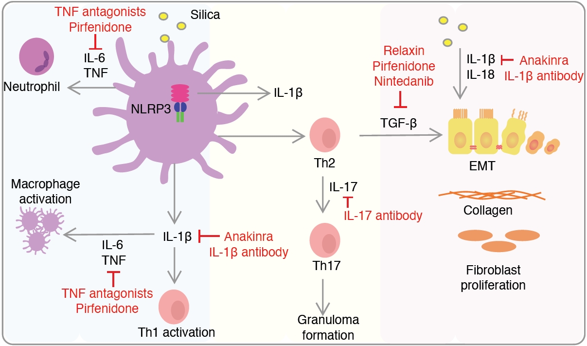
Mechanisms of inflammatory and fibrotic pathways in silicosis. Silica uptake from macrophages activates the nucleotide-binding domain, leucine-rich–containing family, pyrin domain–containing-3 (NLRP3) inflammasome and induces nuclear factor кB activation and the production of cytokines such as interleukin (IL)-6, tumor necrosis factor (TNF), and pro–IL-1β itself, as well as the migration of type 1 alveolar macrophages and neutrophils to the airways. These inflammatory cells attract Th1 lymphocytes, which in turn secrete interferon γ and TNF. Fibrosis is driven by the pro-fibrotic mediator transforming growth factor beta (TGF-β). TGF-β and IL-6 contribute to the differentiation of Th2 to Th17 lymphocytes, which promotes the formation of granulomas via the production of IL-17. Silica increases the production of IL-1β and IL-18 and induces epithelial-mesenchymal transition (EMT), where epithelial cells acquire a more mesenchymal-like phenotype. EMT promotes further synthesis and deposition of collagen. Sites of actions of compounds studied in silicosis are depicted. Adapted from Barnes et al. Curr Opin Pulm Med 2024;30(2):185-94, with permission of Wolters Kluwer Health, Inc.11
Fig. 2.
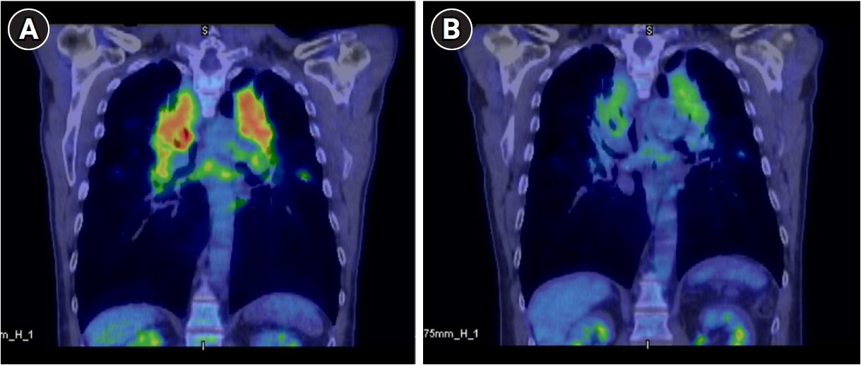
Positron emission tomography scans pretreatment (A) and post-treatment (B). Red indicates markedly increased fluorine-18 fluorodeoxyglucose uptake.
Fig. 3.
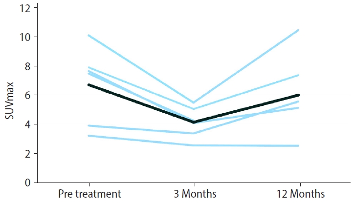
Maximum standardized uptake value (SUVmax) at baseline, three months post-treatment, and 12 months post-treatment. Individual patient data is depicted in light blue, and the average in dark blue.
Table 1.
Participant baseline characteristics
| Variable | Value |
|---|---|
| Male sex | 7 (100) |
| Age at diagnosis (years) | 45 (37–49) |
| Duration of silica exposure (years) | 19 (15–21) |
| Time from last exposure (years) | 4 (1–11) |
Table 2.
Change in outcome measures at baseline and 3 months post-prednisolone
| Variable | Pre | Post | Mean change (post − pre) | p-value |
|---|---|---|---|---|
| PET parameters | ||||
| SUVmaxa | 6.7 ± 2.6 | 4.2 ± 1.0 | –2.5 ± 1.5 | 0.012 |
| SUVmeana | 1.2 ± 0.5 | 1.0 ± 0.3 | –0.2 ± 0.4 | 0.107 |
| % Lung >1 SUVa | 49.7 ± 36.4 | 45.8 ± 28.4 | –3.9 ± 5.7 | 0.527 |
| % Lung >2.5 SUVa | 7.0 ± 6.3 | 2.4 ± 2.2 | –4.6 ± 1.7 | 0.038 |
| TLGa | 4,065 ± 1,535 | 3,629 ± 1,113 | –436 ± 711 | 0.187 |
| Pulmonary function tests | ||||
| FEV1 (L) | 2.5 ± 1.1 | 2.4 ± 1.1 | –0.1 ± 0.2 | 0.328 |
| FEV1 % predicted | 65.3 ± 26.6 | 63 ± 26 | –2.3 ± 4.9 | 0.265 |
| FVC (L) | 4.1 ± 0.8 | 4 ± 0.8 | –0.1 ± 0.2 | 0.237 |
| FVC % predicted | 85.9 ± 11.7 | 83.7 ± 13.2 | –2.1 ± 4.5 | 0.253 |
| DLCO (mL/min/mmHg) | 20.7 ± 6.5 | 21.6 ± 7.1 | 0.9 ± 2.2 | 0.314 |
| DLCO % predicted | 72.4 ± 19.7 | 70.7 ± 23.6 | –1.7 ± 7.5 | 0.567 |
| Serum inflammatory markers | ||||
| ACE (U/L) | 95 ± 45.7 | 65.2 ± 35.2 | –29.8 ± 11.7 | 0.005 |
| ESR (mm/h) | 13.4 ± 7.8 | 15.6 ± 24 | 2.2 ± 21.1 | 0.827 |
| Quality of life | ||||
| SGRQ total scoreb | 41.8 ± 24.8 | 46.7 ± 23.7 | 4.87 ± 15.3 | 0.570 |
| SGRQ symptomsb | 53.1 ± 31.4 | 62.8 ± 34.9 | 9.76 ± 19.9 | 0.400 |
| SGRQ activityb | 48.8 ± 22.1 | 39.2 ± 10.8 | –9.6 ± 24.3 | 0.487 |
| SGRQ impactb | 33.8 ± 25.1 | 45.5 ± 31.7 | 11.7 ± 14.2 | 0.198 |
| ICOERD domains | ||||
| Well defined opacity profusion score | 7 ± 6.6 | 6 ± 5.7 | –1 ± 1.6 | 0.058 |
| Irregular linear opacity profusion score | 3 ± 1.8 | 3.4 ± 2.2 | 0.4 ± 2.2 | 0.629 |
| Large opacities | 2.6 ± 1.8 | 2.3 ± 1.7 | –0.3 ± 0.5 | 0.172 |
Values are presented as mean ± SD.
PET: positron emission tomography; SUV: standardized uptake value; TLG: total lung glycolysis; FEV1: forced expiratory volume in one second; FVC: forced vital capacity; DLCO: diffusing capacity for carbon monoxide; ACE: angiotensin-converting enzyme; ESR: erythrocyte sedimentation rate; SGRQ: St. George's Respiratory Questionnaire; ICOERD: International Classification of High-resolution Computed Tomography for Occupational and Environmental Respiratory Diseases; SD: standard deviation.
Total participants = 7 unless otherwise indicated:
an = 6,
bn = 4.
- 1. Hoy RF, Dimitriadis C, Abramson M, Glass DC, Gwini S, Hore-Lacy F, et al. Prevalence and risk factors for silicosis among a large cohort of stone benchtop industry workers. Occup Environ Med 2023;80(8):439–46.ArticlePubMedPMC
- 2. Leon-Jimenez A, Hidalgo-Molina A, Conde-Sanchez MA, Perez-Alonso A, Morales-Morales JM, Garcia-Gamez EM, et al. Artificial stone silicosis: rapid progression following exposure cessation. Chest 2020;158(3):1060–8.ArticlePubMed
- 3. Kramer MR, Blanc PD, Fireman E, Amital A, Guber A, Rhahman NA, et al. Artificial stone silicosis [corrected]: disease resurgence among artificial stone workers. Chest 2012;142(2):419–24.ArticlePubMed
- 4. Fazio JC, Gandhi SA, Flattery J, Heinzerling A, Kamangar N, Afif N, et al. Silicosis among immigrant engineered stone (Quartz) countertop fabrication workers in California. JAMA Intern Med 2023;183(9):991–8.ArticlePubMedPMC
- 5. Yang H, Yang L, Zhang J, Chen J. Natural course of silicosis in dust-exposed workers. J Huazhong Univ Sci Technolog Med Sci 2006;26(2):257–60.ArticlePubMedPDF
- 6. Zheng D, Liwinski T, Elinav E. Inflammasome activation and regulation: toward a better understanding of complex mechanisms. Cell Discov 2020;6:36.ArticlePubMedPMCPDF
- 7. Song L, Weng D, Dai W, Tang W, Chen S, Li C, et al. Th17 can regulate silica‐induced lung inflammation through an IL‐1β‐dependent mechanism. J Cell Mol Med 2014;18(9):1773–84.ArticlePubMedPMCPDF
- 8. Ramkissoon C, Gaskin S, Hall T, Pisaniello D, Zosky G. Engineered stone fabrication work releases volatile organic compounds classified as lung irritants. Ann Work Expo Health 2023;67(2):288–93.ArticlePubMedPMCPDF
- 9. Barnes H, Goh NS, Leong TL, Hoy R. Silica-associated lung disease: an old-world exposure in modern industries. Respirology 2019;24(12):1165–75.ArticlePubMedPDF
- 10. Leon-Jimenez A, Rodriguez-Rubio Corona J, Jimenez-Gomez G, Pinero Fernandez-Reyes ML, Hidalgo-Molina A, Pajares-Vinardel M, et al. High metabolic activity in positron emission tomography and systemic inflammation occurring years after exposure cessation in engineered stone silicosis. Sci Rep 2025;15(1):25364.ArticlePubMedPMC
- 11. Barnes H, Lam M, Tate MD, Hoy R. Toward targeted treatments for silicosis. Curr Opin Pulm Med 2024;30(2):185–94.ArticlePubMed
- 12. Franchimont D. Overview of the actions of glucocorticoids on the immune response: a good model to characterize new pathways of immunosuppression for new treatment strategies. Ann N Y Acad Sci 2004;1024:124–37.ArticlePubMed
- 13. Ayroldi E, Cannarile L, Migliorati G, Nocentini G, Delfino DV, Riccardi C. Mechanisms of the anti-inflammatory effects of glucocorticoids: genomic and nongenomic interference with MAPK signaling pathways. FASEB J 2012;26(12):4805–20.ArticlePubMedPDF
- 14. Liberman AC, Budzinski ML, Sokn C, Gobbini RP, Steininger A, Arzt E. Regulatory and mechanistic actions of glucocorticoids on T and inflammatory cells. Front Endocrinol (Lausanne) 2018;9:235.ArticlePubMedPMC
- 15. Baughman RP, Valeyre D, Korsten P, Mathioudakis AG, Wuyts WA, Wells A, et al. ERS clinical practice guidelines on treatment of sarcoidosis. Eur Respir J 2021;58(6):2004079.ArticlePubMedPMC
- 16. Zincirkeser S, Sahin E, Halac M, Sager S. Standardized uptake values of normal organs on 18F-fluorodeoxyglucose positron emission tomography and computed tomography imaging. J Int Med Res 2007;35(2):231–6.ArticlePubMedPDF
- 17. Yoon HY, Lee SH, Ha S, Ryu JS, Song JW. The value of (18)F-FDG PET/CT in evaluating disease severity and prognosis in idiopathic pulmonary fibrosis patients. J Korean Med Sci 2021;36(41):e257.ArticlePubMedPMCPDF
- 18. Culver BH, Graham BL, Coates AL, Wanger J, Berry CE, Clarke PK, et al. Recommendations for a standardized pulmonary function report: an official American Thoracic Society technical statement. Am J Respir Crit Care Med 2017;196(11):1463–72.ArticlePubMedPDF
- 19. Liu H, Yan B, Han B, Sun J, Yang Y, Chen J. Assessment of respiration-related quality of life of Chinese patients with silicosis and its influencing factors using the St. George's Respiratory Questionnaire (SGRQ). J Clin Nurs 2012;21(11-12):1515–23.ArticlePubMed
- 20. Souza TP, Souza R, Watte G, de Souza JA, Moreira JD, Knorst MM. Predictors of health-related quality of life among semi-precious stone mineworkers exposed to silica dust. Int Arch Occup Environ Health 2021;94(4):679–87.ArticlePubMedPDF
- 21. Hoy RF, Glass DC, Dimitriadis C, Hansen J, Hore-Lacy F, Sim MR. Identification of early-stage silicosis through health screening of stone benchtop industry workers in Victoria, Australia. Occup Environ Med 2021;78(4):296–302.ArticlePubMed
- 22. Des Jarlais DC, Lyles C, Crepaz N; TREND Group. Improving the reporting quality of nonrandomized evaluations of behavioral and public health interventions: the TREND statement. Am J Public Health 2004;94(3):361–6.ArticlePubMedPMC
- 23. Levin K, McLean C, Hoy R. Artificial stone-associated silicosis: clinical-pathological-radiological correlates of disease. Respirol Case Rep 2019;7(7):e00470.ArticlePubMedPMCPDF
- 24. Go LH, Rose CS, Zell-Baran LM, Almberg KS, Iwaniuk C, Clingerman S, et al. Historical shift in pathological type of progressive massive fibrosis among coal miners in the USA. Occup Environ Med 2023;80(8):425–30.ArticlePubMedPMC
- 25. Sharma SK, Pande JN, Verma K. Effect of prednisolone treatment in chronic silicosis. Am Rev Respir Dis 1991;143(4 Pt 1):814–21.ArticlePubMedPDF
- 26. Hua JT, Cool CD, Fireman Klein E, Adir Y, Lee LJ, Zell-Baran LM, et al. Silicosarcoidosis: histologic and clinical features of an occupational granulomatous disease. Am J Ind Med 2025;68(6):491–507.ArticlePubMedPMC
- 27. Ronsmans S, Nemery B, De Crem N, Weynand B. Silicosis, sarcoidosis, or both? Rethinking disease labels in light of co-occurrence. Am J Ind Med 2025;68(6):487–90.ArticlePubMed
- 28. Dufresne A, Begin R, Dion C, Jagirdar J, Rom WN, Loosereewanich P, et al. Angular and fibrous particles in lung in relation to silica-induced diseases. Int Arch Occup Environ Health 1998;71(4):263–9.ArticlePubMedPDF
- 29. Huntley CC, Burge PS, Moore VC, Robertson AS, Walters GI. Pulmonary function decline and survival in silicosis: a retrospective longitudinal study. Thorax 2022;77:A6–7.
REFERENCES
Figure & Data
REFERENCES
Citations
Citations to this article as recorded by 
 Cite
Cite XML Download
Evaluating the efficacy of prednisolone for silicosis with progressive massive fibrosis in Australia: a pre-post quasi-experimental study
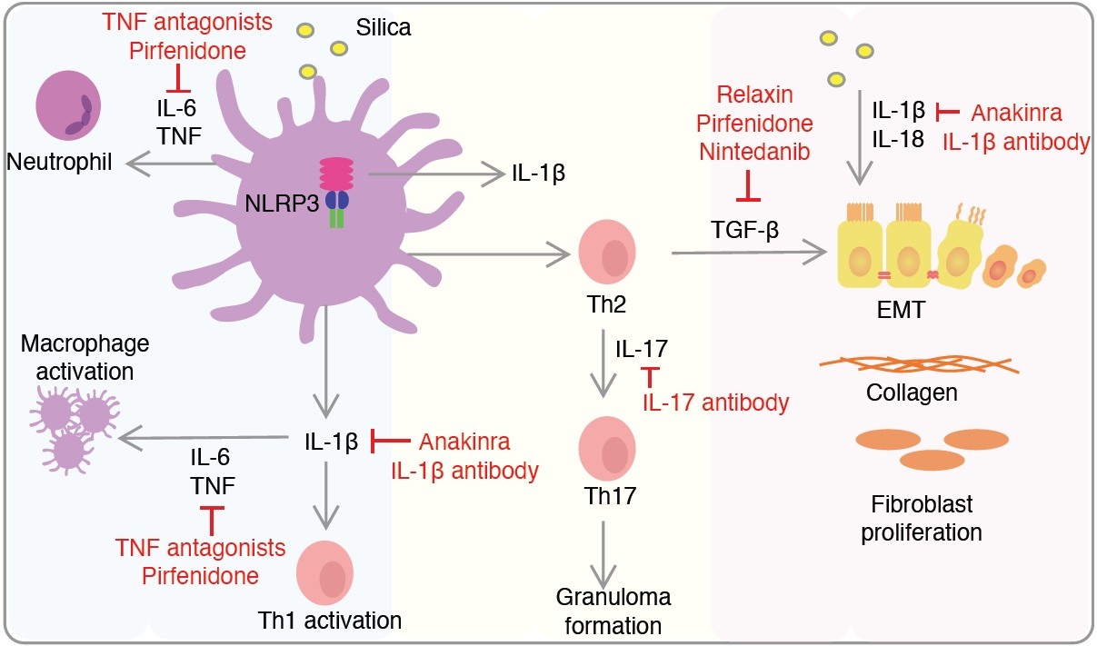
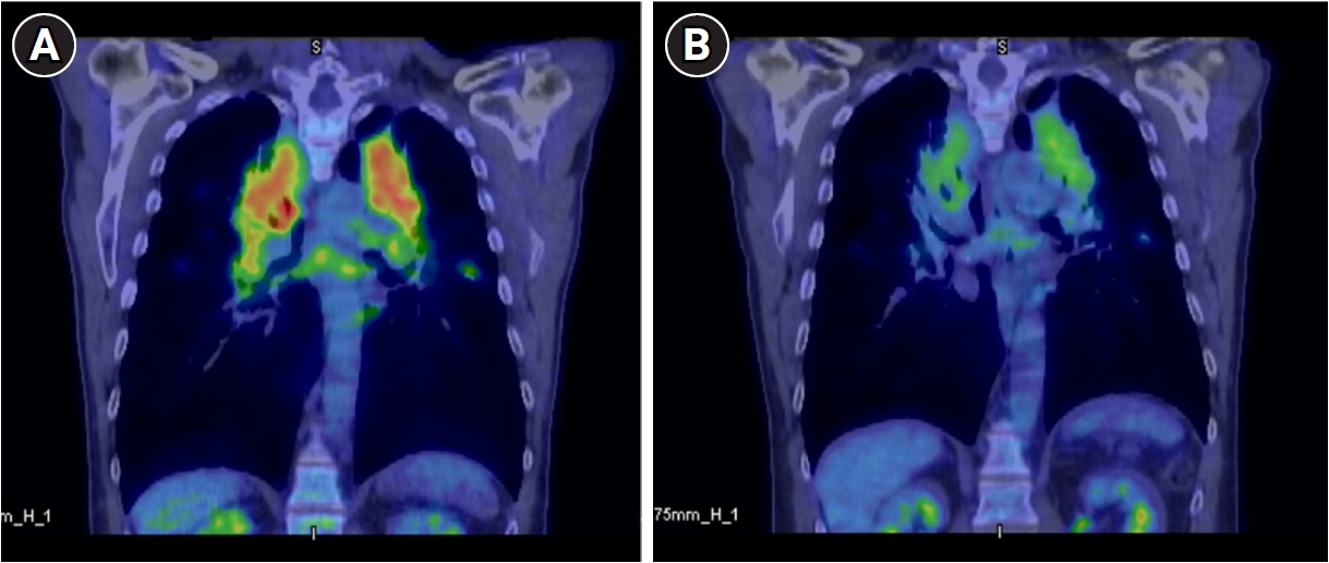
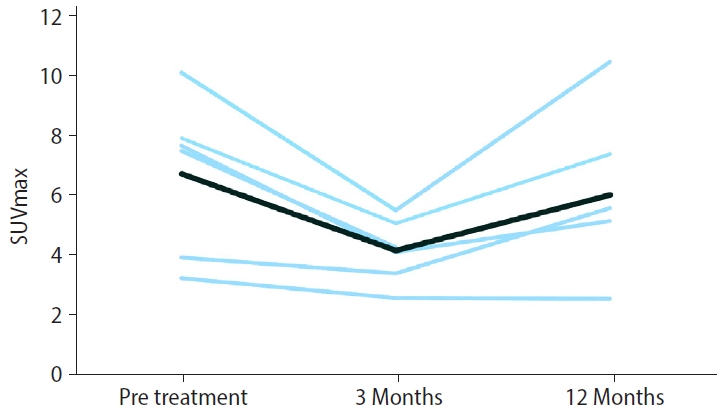
Fig. 1. Mechanisms of inflammatory and fibrotic pathways in silicosis. Silica uptake from macrophages activates the nucleotide-binding domain, leucine-rich–containing family, pyrin domain–containing-3 (NLRP3) inflammasome and induces nuclear factor кB activation and the production of cytokines such as interleukin (IL)-6, tumor necrosis factor (TNF), and pro–IL-1β itself, as well as the migration of type 1 alveolar macrophages and neutrophils to the airways. These inflammatory cells attract Th1 lymphocytes, which in turn secrete interferon γ and TNF. Fibrosis is driven by the pro-fibrotic mediator transforming growth factor beta (TGF-β). TGF-β and IL-6 contribute to the differentiation of Th2 to Th17 lymphocytes, which promotes the formation of granulomas via the production of IL-17. Silica increases the production of IL-1β and IL-18 and induces epithelial-mesenchymal transition (EMT), where epithelial cells acquire a more mesenchymal-like phenotype. EMT promotes further synthesis and deposition of collagen. Sites of actions of compounds studied in silicosis are depicted. Adapted from Barnes et al. Curr Opin Pulm Med 2024;30(2):185-94, with permission of Wolters Kluwer Health, Inc.11
Fig. 2. Positron emission tomography scans pretreatment (A) and post-treatment (B). Red indicates markedly increased fluorine-18 fluorodeoxyglucose uptake.
Fig. 3. Maximum standardized uptake value (SUVmax) at baseline, three months post-treatment, and 12 months post-treatment. Individual patient data is depicted in light blue, and the average in dark blue.
Fig. 1.
Fig. 2.
Fig. 3.
Evaluating the efficacy of prednisolone for silicosis with progressive massive fibrosis in Australia: a pre-post quasi-experimental study
| Variable | Value |
|---|---|
| Male sex | 7 (100) |
| Age at diagnosis (years) | 45 (37–49) |
| Duration of silica exposure (years) | 19 (15–21) |
| Time from last exposure (years) | 4 (1–11) |
| Variable | Pre | Post | Mean change (post − pre) | p-value |
|---|---|---|---|---|
| PET parameters | ||||
| SUVmax |
6.7 ± 2.6 | 4.2 ± 1.0 | –2.5 ± 1.5 | 0.012 |
| SUVmean |
1.2 ± 0.5 | 1.0 ± 0.3 | –0.2 ± 0.4 | 0.107 |
| % Lung >1 SUV |
49.7 ± 36.4 | 45.8 ± 28.4 | –3.9 ± 5.7 | 0.527 |
| % Lung >2.5 SUV |
7.0 ± 6.3 | 2.4 ± 2.2 | –4.6 ± 1.7 | 0.038 |
| TLG |
4,065 ± 1,535 | 3,629 ± 1,113 | –436 ± 711 | 0.187 |
| Pulmonary function tests | ||||
| FEV1 (L) | 2.5 ± 1.1 | 2.4 ± 1.1 | –0.1 ± 0.2 | 0.328 |
| FEV1 % predicted | 65.3 ± 26.6 | 63 ± 26 | –2.3 ± 4.9 | 0.265 |
| FVC (L) | 4.1 ± 0.8 | 4 ± 0.8 | –0.1 ± 0.2 | 0.237 |
| FVC % predicted | 85.9 ± 11.7 | 83.7 ± 13.2 | –2.1 ± 4.5 | 0.253 |
| DLCO (mL/min/mmHg) | 20.7 ± 6.5 | 21.6 ± 7.1 | 0.9 ± 2.2 | 0.314 |
| DLCO % predicted | 72.4 ± 19.7 | 70.7 ± 23.6 | –1.7 ± 7.5 | 0.567 |
| Serum inflammatory markers | ||||
| ACE (U/L) | 95 ± 45.7 | 65.2 ± 35.2 | –29.8 ± 11.7 | 0.005 |
| ESR (mm/h) | 13.4 ± 7.8 | 15.6 ± 24 | 2.2 ± 21.1 | 0.827 |
| Quality of life | ||||
| SGRQ total score |
41.8 ± 24.8 | 46.7 ± 23.7 | 4.87 ± 15.3 | 0.570 |
| SGRQ symptoms |
53.1 ± 31.4 | 62.8 ± 34.9 | 9.76 ± 19.9 | 0.400 |
| SGRQ activity |
48.8 ± 22.1 | 39.2 ± 10.8 | –9.6 ± 24.3 | 0.487 |
| SGRQ impact |
33.8 ± 25.1 | 45.5 ± 31.7 | 11.7 ± 14.2 | 0.198 |
| ICOERD domains | ||||
| Well defined opacity profusion score | 7 ± 6.6 | 6 ± 5.7 | –1 ± 1.6 | 0.058 |
| Irregular linear opacity profusion score | 3 ± 1.8 | 3.4 ± 2.2 | 0.4 ± 2.2 | 0.629 |
| Large opacities | 2.6 ± 1.8 | 2.3 ± 1.7 | –0.3 ± 0.5 | 0.172 |
Table 1. Participant baseline characteristics
Values are presented as number (%) or mean (range).
Table 2. Change in outcome measures at baseline and 3 months post-prednisolone
Values are presented as mean ± SD. PET: positron emission tomography; SUV: standardized uptake value; TLG: total lung glycolysis; FEV1: forced expiratory volume in one second; FVC: forced vital capacity; DLCO: diffusing capacity for carbon monoxide; ACE: angiotensin-converting enzyme; ESR: erythrocyte sedimentation rate; SGRQ: St. George's Respiratory Questionnaire; ICOERD: International Classification of High-resolution Computed Tomography for Occupational and Environmental Respiratory Diseases; SD: standard deviation. Total participants = 7 unless otherwise indicated: n = 6, n = 4.


