 KSOEM
KSOEM

Articles
- Page Path
- HOME > Ann Occup Environ Med > Volume 38; 2026 > Article
- Case Report Sinonasal hemangiosarcoma following juvenile occupational exposure to formaldehyde when breeding farm animals
-
Daniela Napolitano1,2
 , Stefania Massacesi1,2, Roberto Calisti1,2,*
 -
Annals of Occupational and Environmental Medicine 2026;38:e27.
DOI: https://doi.org/10.35371/aoem.2026.38.e27
Published online: July 13, 2026
1Occupational Safety and Health – Occupational Epidemiology Unit, Civitanova Marche, Italy
2Marche Regional Operation Centre (COR) of the National Registry of Sino-Nasal Cancers (ReNaTuNS), Civitanova Marche, Italy
- *Corresponding author: Roberto Calisti Occupational Safety and Health – Occupational Epidemiology Unit, Civitanova Marche, Macerata 62012, Macerata, Italy E-mail: roberto.calisti.5742@gmail.com
• Received: February 27, 2026 • Revised: July 1, 2026 • Accepted: July 2, 2026
© 2026 Korean Society of Occupational & Environmental Medicine
This is an Open Access article distributed under the terms of the Creative Commons Attribution Non-Commercial License (https://creativecommons.org/licenses/by-nc/4.0/) which permits unrestricted non-commercial use, distribution, and reproduction in any medium, provided the original work is properly cited.
- 877 Views
- 30 Download


Abstract
-
Background Hemangiosarcomas are heterogeneous malignancies possibly originating in any part of the body, rare in humans. The web of causation of human hemangiosarcomas remains poorly understood, to date, except for subsets of cases which have been recognized in a well-defined causal correlation to vinyl chloride or to ionizing radiation. In 2005, the International Agency for Research on Cancer ranked formaldehyde as a human carcinogen, causally related to both nasopharyngeal cancers and acute non-lymphocytic leukemias; at the time, the evidence in favor of a causal relationship of formaldehyde to sinonasal cancers was stated as “limited”. In 2024, an umbrella review reported a significantly increased risk for sinonasal cancers among formaldehyde-exposed workers.
-
Case presentation A sinonasal hemangiosarcoma affecting an Italian male born in 1957, diagnosed in 2022, was reported to the Italian National Registry of Sino-Nasal Cancers (ReNaTuNS); the patient’s occupational history revealed a remote exposure to formaldehyde, used as a disinfectant, when breeding farm animals, from 1967 to 1976. No previous reporting has been found about a possible association between formaldehyde and sinonasal non-epithelial cancers.
-
Conclusions Formaldehyde is, in our opinion, a suitable candidate to be considered for playing a causal role in the causation of the neoplasm; formal studies are required to test the hypothesis.
BACKGROUND
Hemangiosarcomas are anatomically and genomically heterogeneous malignancies originating in the endothelial linings of the blood vessels, possibly in any part of the body: they are rare in humans.1 The sinonasal tract is a particularly rare primary localization of human hemangiosarcomas, with just individual cases and a single series of 10 cases retrieved through PubMed from 2000 onwards.2-8 In several animal species, hemangiosarcomas are relatively frequent neoplasms instead; it is worth noting that, in dogs, cats and horses, these tumors often arise from the skin of short-haired, lightly pigmented individuals, reliably correlating to chronic damage from solar radiation.9
The web of causation of the human hemangiosarcomas remains poorly understood, to date, except for subsets of cases which have been recognized in a well-defined causal correlation to vinyl chloride or to ionizing radiation.10-18 Liver hemangiosarcomas arising in workers occupationally exposed to vinyl chloride have been reported since 1974; the majority of them were identified in the United States and in Italy.10-13 Thorotrast, a radiographic contrast medium composed of thorium dioxide (ThO2) and Th-232, medically used from 1930 to 1955, was recognized as the causative agent of liver hemangiosarcomas, arising also decades after the administration of the agent.14-16 Cancer patients receiving radiation therapy could suffer hemangiosarcomas in the radiation field, even after only 3 years following the start of their treatment.17,18 Sixteen liver hemangiosarcomas were diagnosed from 1948 to 2018 in a cohort of 22,377 workers hired between 1948 and 1982 at a Russian nuclear plant, being exposed to gamma rays and alpha particles there.19 No formal epidemiological studies have been retrieved about the causation of the human sinonasal hemangiosarcomas.
In 2005, International Agency for Research on Cancer ranked formaldehyde as a human carcinogen (group 1), which acts through both genotoxic and non-genotoxic mechanisms, and causally correlates to both nasopharyngeal cancers and acute non-lymphocytic leukemias; at the time, the evidence in favor of a causal relationship of formaldehyde to sinonasal cancers was stated as “limited.”20 In 2024, an umbrella review explored the available evidence associating some occupational carcinogens and cancer risk at 19 anatomical sites; a relative risk of 1.68 (95% confidence interval: 1.38–2.05) was reported for sinonasal cancers among the subjects having been occupationally exposed to formaldehyde.21
It is worth noting that, to date, the body of evidence about the etiology of sinonasal cancers almost entirely refers to malignancies of epithelial origin; namely, no previous reporting has been found about an association between formaldehyde and human non-epithelial malignancies arising in the sinonasal tract.
ReNaTuNS (“Registro Nazionale dei Tumori Naso-Sinusali”) is a specialized cancer registry established in Italy by law in 2008 (Article 244 of the Legislative Decree 81/08).
ReNaTuNS, based on a network of Regional Operation Centres (“Centri Operativi Regionali” - COR), systematically identifies all the sinonasal cancers of epithelial origin arising in the general Italian population, collects detailed anamnestic data on the basis of a standardized questionnaire, and evaluates the individual profiles of exposure to established carcinogens and other possibly relevant agents; it also publishes epidemiological analyses of the case series.22
Our Unit, one of the aforementioned ReNaTuNS CORs, identified and studied around 320 cases of epithelial sinonasal cancers which arose from 1998 onwards in a local population of roughly 1.5 million; cases of non-epithelial sinonasal cancer have also been occasionally detected and studied.
CASE PRESENTATION
A sinonasal hemangiosarcoma was reported by a general practitioner to ReNaTuNS; even though this very particular neoplasm of non-epithelial origin failed to meet the current ReNaTuNS inclusion criteria, we decided to investigate it as a possible sentinel event. No previous reporting had been found about a possible association between formaldehyde and sinonasal non-epithelial cancers. The patient consented to be interviewed and the ReNaTuNS standardized questionnaire was administered. The patient’s informed consent to the possible publication of a case report was acquired at a later time.
The patient, an Italian male born in 1957, was referred in 2019 for long-lasting nasal obstruction that, in March 2022, was complicated by a sudden emission of abundant, foul-smelling fluid. In April 2022, a computed tomography (CT) revealed a solid mass, about 5 cm across, occupying the right maxillary and ethmoidal sinuses (Fig. 1). In May 2022, the patient underwent a functional endoscopic sinus surgery at a local hospital. A local pathologist histologically diagnosed a hemangiosarcoma; he described, in a context of necrosis and suppuration, clusters of large, pleomorphic epithelioid cancer cells, showing irregularly profiled nuclei and prominent nucleoli, testing positive for ERG and CD31, negative for AE1/AE3, S100, muscle-specific actin - clone HHF35, and desmin. In August 2022, at a specialized cancer center, the available histological material was reviewed by a second pathologist. He confirmed the diagnosis, describing a vessel-forming neoplasm combining newly formed vessels lined by atypical endothelial cells and solid areas of epithelioid cells with prominent nucleoli; the cancer cells tested positive for ERG, CD31, CD34, c-Myc (rarely), and MIB (Ki-67) (rarely), negative for p53 (DO7), and smooth muscle actin - clone 1A4.
In October 2022, a magnetic resonance (MR) recorded the presence of residual cancer tissue along the margin of the surgical resection (Fig. 2). The cancer center proposed a further, largely destructive, intervention flanked by adjuvant chemotherapy; the patient refused. Between December 2022 and February 2023, a proton therapy (33 sessions) was tentatively carried out.
In October 2025, an MR recorded no presence of residual cancer (Fig. 3). Even later, over 36 months after the end of the radiotherapy, no evidence of cancer recurrence emerged; the patient is thus experiencing a cancer-free period much longer than expected.1,23
The patient suffered skin scars in the radiation field, a pronounced reduction of the right-eye vision, and a continuous, abundant, bilateral lacrimation due to obstruction of the nasolacrimal ducts. The visual loss and the lacrimation make walking difficult and driving vehicles impossible, but the patient considers the quality of his life dignified. No previous relevant head and neck diseases and no previous radiotherapy were reported by the patient.
The patient, a never smoker, held just two jobs. From 1967 to 1976 (between 10 and 19 years of age), he was a farmer and a breeder on the family farm, where cereals, sugar beets, vegetables, wine vines and olive trees were grown and cattle, poultry and rabbits were raised. Throughout this period, he regularly disinfected the livestock stables by means of water-based formaldehyde solutions, sprinkled onto the surfaces (not directly onto the animals) by a hand pump backpack sprayer. This kind of device basically consists of a tank containing the solution to be sprayed and carried on the worker’s back by two padded shoulder straps, a manually operated pump, and a pipe system which takes the pressurized fluid to the spraying lance. While preparing and spraying the disinfectant, the subject never wore respiratory protection or other protective equipment.
He also prepared and sprayed fungicide solutions, namely “verdigris” and other unspecified “sulfur products”, on crops and trees; due to their strong, bad smell, on those occasions he wore a tied kerchief to cover his mouth and nose, as the only personal protection. From time to time, pest control operators from the local municipality prepared and sprinkled unspecified insecticides inside the stables, also in the presence of the subject; even on these occasions, he never wore respiratory protection or other protective equipment.
The approximately 9 years of work on the family farm were followed by a 25-year-long career (1977–2002, i.e., between 20 and 45 years of age) as a “Carabiniere”, i.e., a trooper in an Italian national military in charge of territorial control police duties. He was assigned to different garrisons in Italy, in the absence of any involvement in special operations within the country or abroad. The subject denied having wiped or lubricated weapons at any time during this period, as these tasks were reserved for specialized armorers. After his early retirement, in 2002 (when he was 45 years of age), the subject did not engage in any further work activity.
No clue of exposure to vinyl chloride and ionizing radiation emerges from the patient’s history (Thorotrast was no longer in use before the patient was born). As a farmer and breeder, the patient was certainly exposed to a set of airborne chemicals, i.e., “verdigris” (a fungicidal mixture of copper sulfate and calcium hydroxide), other fungicidal “sulfur products,” some not better identified insecticides, and formaldehyde; this topic will be covered further. Due to the patient’s denial of having wiped and lubricated weapons, no exposure to organic solvents, possibly containing benzene, or to mineral oils, possibly containing polycyclic aromatic hydrocarbons, can be speculated.
No extra-work exposure to possibly relevant agents emerged from the interview; the fact that the patient was never a smoker is worthy of consideration.
As the recognition of all cancers of possible occupational origin is mandatory in Italy, a “case by case” ethical approval of such investigations is not required. Nevertheless, our Unit always requests informed consent from the patients whose individual cases are proposed for scientific publication or educational discussion. Written informed consent was obtained from the patient for the publication of this case report and any accompanying clinical data. The patient himself produced a short text describing his personal experience of illness and medical treatment, authorizing its publication too, if required.
DISCUSSION AND CONCLUSION
The patient began to perceive long-lasting nasal obstruction when he was 62 years old and suffered a sudden emission of abundant, foul-smelling fluid when he was 65 years old. He was diagnosed with sinonasal hemangiosarcoma, showing an epithelioid phenotype, on the basis of an initial histological examination and immunohistochemistry performed two months after the above-mentioned incident, subsequently confirmed. Epithelioid hemangiosarcoma constitutes a defined, particularly rare subtype within the angiosarcoma spectrum, typically unresponsive to radiation therapy; the present case is peculiar also in this regard.23 Why did the patient contract the disease?
We consider that the 9 years the patient spent on the family farm are the only ones of interest in his occupational history. “Verdigris” and “sulfur products” are generally recognized respiratory irritants, but no suggestion of a cancer risk has been currently associated with them. Several insecticides are established carcinogens, but the lack of specific information about the formulation of every preparation the patient was exposed to makes it impossible to neglect or claim a pathogenic contribution from these agents; in any case, if it had been present, nothing would exclude the possibility that also other agents had entered the web of causation of the disease. Airborne formaldehyde is definitely present in the patient’s occupational experience. Considering that he personally prepared the disinfectant solution and sprayed it inside confined spaces, in the absence of any personal protection, while acknowledging the limitations of reconstructing exposures that occurred several decades ago, we believe that the exposure to the agent would have reached relevant peak levels. Years ago, having performed a set of short-time samplings in various Italian work scenarios, our group found indeed high levels of formaldehyde vapor in stables where formaldehyde was in use as a disinfectant, in the absence of specific measures to reduce the air spreading of the agent, recording a peak level of 3,669.0 μg/m3 in a poultry farm.24,25
But did formaldehyde play a role in the web of causation of the investigated malignancy or are we dealing with a mere, non-causal association? This question cannot receive an ultimate answer at present, but we believe that some topics can be usefully discussed. An interval of 55 years elapsed from the beginning of the exposure to formaldehyde and the diagnosis, and an interval of 46 years elapsed from the discontinuation of the exposure to formaldehyde and the diagnosis (Table 1); these are really long latency periods, but they do not appear extraordinary in occupational carcinogenesis.
Why is the epidemiological evidence of formaldehyde’s carcinogenicity in humans strong for nasopharyngeal cancers, although less so for sinonasal cancers? This apparent inconsistency might be explained, at least in part, by the fact that frequently, in the epidemiological studies, the nasal tumors are jointly classified with the tumors of the nasal sinuses. It has been pointed out that formaldehyde vapor is largely absorbed in the upper airways, as a whole, but at the same atmospheric concentration, the sinuses receive much less exposure in comparison to the nasal cavities and the nasopharynx.26
A systematic literature review has also indicated formaldehyde as one of the agents most strongly associated with salivary gland tumors.27 A genome-wide mapping of formaldehyde-induced DNA-protein crosslinks has been performed by means of a human fibrosarcoma cell line, providing clues about the pathways of formaldehyde-induced carcinogenesis, not limiting the perspective to only the epithelial tissues.28 Two recent studies have led towards a widening of the spectrum of cancers causally related to formaldehyde to non-epithelial malignancies such as glioblastomas and lymphomas.29,30
Given this, formaldehyde is, in our opinion, a suitable candidate to be considered, having played a role in the genesis of the investigated sinonasal hemangiosarcoma. This hypothesis certainly remains, in the light of the available evidence, nothing but a suggestion; formal investigations would be needed to test it, taking into account that the web of causation of the rare malignancies is particularly difficult to explore.
Specifically designed case-control studies are extremely helpful in this field, but they need a very wide base of study population to be powerful enough to achieve conclusive results; they are therefore burdensome to project and manage.
Dedicated Cancer Registries, systematically integrating an appropriate nosographic definition, the collection of detailed anamnestic data and the assignment of a detailed exposure profile for every enrolled patient, could represent a viable and sustainable tool to approach the study of rare malignancies, such as the sinonasal non-epithelial ones and others.
Abbreviations
COR
Centro Operativo Regionale (Regional Operation Centre)
CT
computed tomography
MR
magnetic resonance
ReNaTuNS
Registro Nazionale dei Tumori Naso-Sinusali (National Registry of Sino-Nasal Cancers)
-
Competing interests
The authors declare that they have no competing interests.
-
Author contributions
Conceptualization: Calisti R, Napolitano D, Massacesi S. Data curation: Massacesi S (who personally takes care of the whole regional section of the ReNaTuNS), Napolitano D (who personally collected the anamnestic data from the patient). Writing - original draft: Calisti R, Massacesi S, Napolitano D. Writing - review and editing: Calisti R, Massacesi S, Napolitano D.
-
Acknowledgments
The authors are very grateful to Dr. Elvira Angeloni, the general practitioner who reported the case to the Italian National Registry of Sino-Nasal Cancers, and to Dr. Roberto De Manno, the specialist radiologist who has reviewed the CT and MR images.
NOTES
Fig. 1.
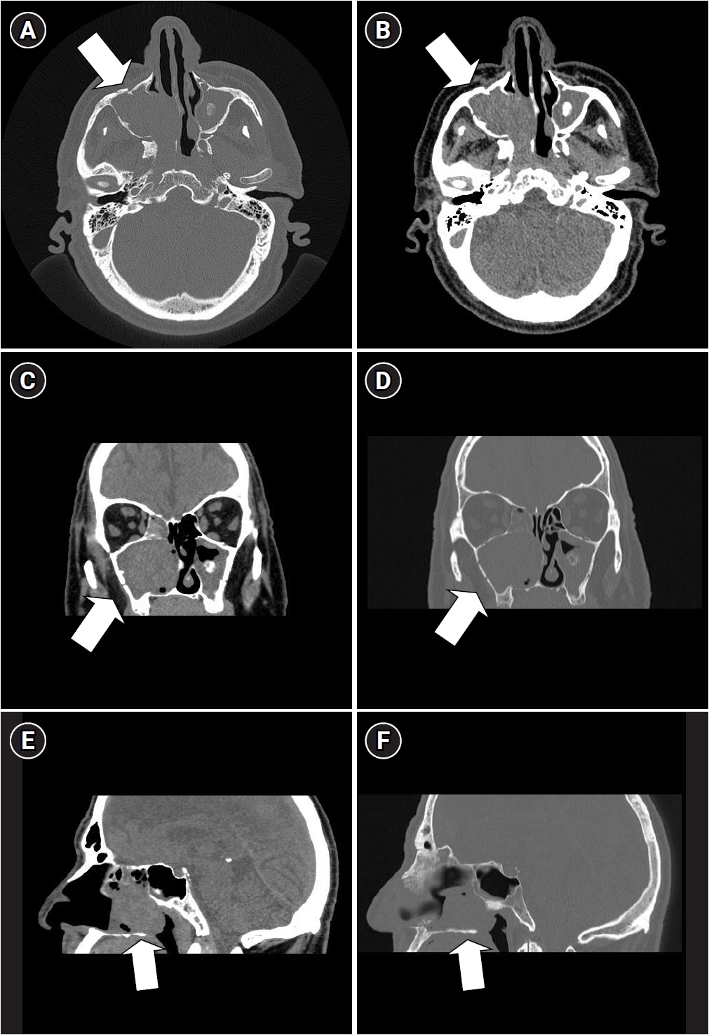
Head computed tomography obtained on 13 Apr 2022. Axial (A, B), coronal (C, D), and sagittal (E, F) images showing an expansive lesion (about 5 cm across), occupying the right nasal-choanal cavity, the right ethmoidal cells and the right maxillary sinus, and eroding the medial sinus wall; evidence of an inflammatory process involving the left ethmoidal cells and the left maxillary sinus. The arrows point to the expansive lesion.
Fig. 2.
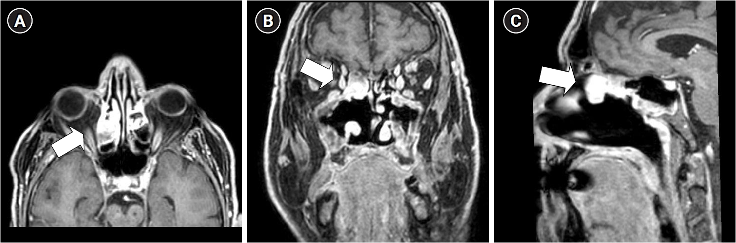
Head magnetic resonance obtained on 10 Oct 2022. Contrast-enhanced axial (A), coronal (B), and sagittal (C) images (paramagnetic contrast medium), showing, on the right side, the persistence of a residual neoplastic lesion expanding towards the upper ethmoidal cells. The arrows point to the expansive lesion.
Fig. 3.
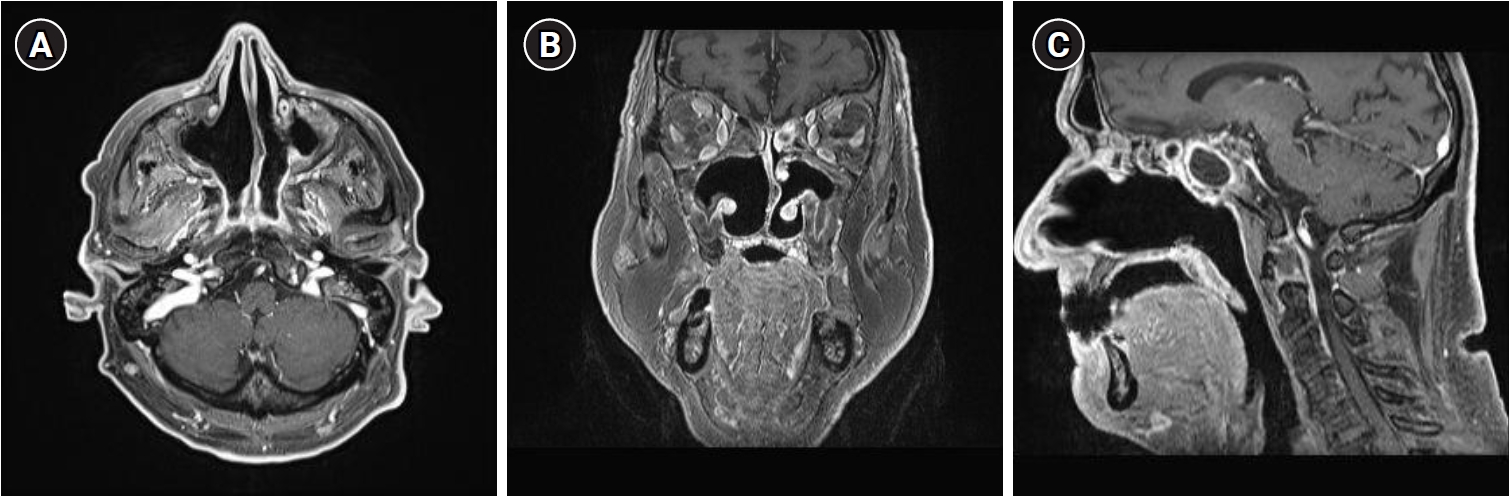
Head magnetic resonance obtained on 1 Oct 2025. Contrast-enhanced axial (A), coronal (B), and sagittal (C) images (paramagnetic contrast medium), showing no persistence of residual neoplastic lesions.
Table 1.
Occupational and disease timelines
- 1. Young RJ, Brown NJ, Reed MW, Hughes D, Woll PJ. Angiosarcoma. Lancet Oncol 2010;11(10):983–91.ArticlePubMed
- 2. Velegrakis GA, Panayiotides JG, Skoulakis CE, Papadakis CE, Papadakis DG, Bizakis JG, et al. Angiosarcoma of the maxillary sinus. J Laryngol Otol 2000;114(5):381–4.ArticlePubMed
- 3. Haferkamp C, Pressler H, Koitschev A. Angiosarcoma of the frontal sinus: case report and review of the literature. HNO 2000;48(9):684–8.ArticlePubMedPDF
- 4. Nelson BL, Thompson LD. Sinonasal tract angiosarcoma: a clinicopathologic and immunophenotypic study of 10 cases with a review of the literature. Head Neck Pathol 2007;1(1):1–12.ArticlePubMedPMCPDF
- 5. Chai C, Feng X, Li K, Yan Z, Tan S, Weng J, et al. Paranasal sinus angiosarcoma with facial paralysis as a novel manifestation: a case report and literature review. BMC Neurol 2023;23(1):428.ArticlePubMedPMCPDF
- 6. Narasimhan G, Deshmuk PT, Gaurkar SS, Khan FQ. Angiosarcoma of the maxillary sinus: a case report. Cureus 2024;16(6):e63131.ArticlePubMedPMC
- 7. Kondapaneni R, Florence K, Dooley L, Garrett F. Maxillary sinus angiosarcoma in Cornelia de Lange syndrome: a case report and review of the literature. Cureus 2025;17(4):e82792.ArticlePubMedPMC
- 8. Jakovljevic S, Cvorovic L, Jovanovic K, Kablar D. Nasal angiosarcoma mimicking common sinonasal tumors. J Craniofac Surg 2026;37(7-8):e709–11.ArticlePubMed
- 9. Vilar-Saavedra P, Kitchell BE. Sunlight-induced skin cancer in companion animals. In: Baldi A, Pasquali P, Spugnini EP, editors. Skin Cancer: A Practical Approach. London, UK: Humana Press; 2014, 499–514.
- 10. Editorial: vinyl chloride and cancer. Br Med J 1974;1(5908):590–1.ArticlePubMedPMC
- 11. Maltoni C, Lefemine G, Chieco P, Carretti D. Vinyl chloride carcinogenesis: current results and perspectives. Med Lav 1974;65(11-12):421–44.PubMed
- 12. Pirastu R, Comba P, Reggiani A, Foa V, Masina A, Maltoni C. Mortality from liver disease among Italian vinyl chloride monomer/polyvinyl chloride manufacturers. Am J Ind Med 1990;17(2):155–61.ArticlePubMed
- 13. Fedeli U, Girardi P, Gardiman G, Zara D, Scoizzato L, Ballarin MN, et al. Mortality from liver angiosarcoma, hepatocellular carcinoma, and cirrhosis among vinyl chloride workers. Am J Ind Med 2019;62(1):14–20.ArticlePubMedPDF
- 14. Fruhling L, Gros CM, Batzenschlager A. Generalized angioplastic endothelial sarcoma in a patient 12 years after an intra-arterial and para-arterial thorotrast injection. Bull Assoc Fr Etud Cancer 1955;42(5):559–63.PubMed
- 15. Underwood JC, Huck P. Thorotrast associated hepatic angiosarcoma with 36 years latency. Cancer 1978;42(6):2610–2.ArticlePubMed
- 16. Falk H, Telles NC, Ishak KG, Thomas LB, Popper H. Epidemiology of thorotrast-induced hepatic angiosarcoma in the United States. Environ Res 1979;18(1):65–73.ArticlePubMed
- 17. Anzalone CL, Cohen PR, Diwan AH, Prieto VG. Radiation-induced angiosarcoma. Dermatol Online J 2013;19(1):2.Article
- 18. Ali S, Bains S, Fox E, Desai A, Hallissey M, El-Ghobashy A, et al. Prognostic significance of clinicopathological parameters, margin width and locoregional recurrences on outcome of primary and radiation associated breast angiosarcoma: results from a large UK sarcoma regional service. Breast Cancer Res 2026;28(1):83.ArticlePubMedPMCPDF
- 19. Zhuntova G, Bannikova M, Azizova T. Incidence risk of hepatobiliary malignant neoplasms in the cohort of workers chronically exposed to ionizing radiation. Sci Rep 2024;14(1):17561.ArticlePubMedPMCPDF
- 20. Cogliano VJ, Grosse Y, Baan RA, Straif K, Secretan MB, El Ghissassi F, et al. Meeting report: summary of IARC monographs on formaldehyde, 2-butoxyethanol, and 1-tert-butoxy-2-propanol. Environ Health Perspect 2005;113(9):1205–8.ArticlePubMedPMC
- 21. Xiong X, Zhang S, Liao X, Du J, Zheng W, Hu S, et al. An umbrella review of the evidence associating occupational carcinogens and cancer risk at 19 anatomical sites. Environ Pollut 2024;345:123531.ArticlePubMed
- 22. Marinaccio A, Binazzi A. ReNaTuNS. Sorveglianza Epidemiologica dei Tumori Naso-Sinusali. Manuale Operativo [ReNaTuNS, Epidemiologic Surveillance of the Sinonasal Tumors. Operation Manual]. Roma, Italy: Inail; 2020, 23–6.
- 23. Al Laham O, Sharaf Aldeen R, Ibrahim Basha Z, Ali A, Alhanwt A. Dual unifocal primary epithelioid angiosarcoma: a case report and review of the literature unveiling a rare genre of neoplasia. Int J Surg Case Rep 2024;115:109310.ArticlePubMedPMC
- 24. Calisti R, Isolani L, Mei R. Formaldehyde exposure patterns in a set of Italian indoor workplaces with and without specific emission sources - 2011 - 2018. Ital J Occup Environ Hyg 2018;9(4):165–75.
- 25. Stopponi R, Astuti MC, Mattozzi C. Poultry growers and occupational exposure to formaldehyde. Med Lav 2014;105(3):234.PubMed
- 26. McGregor D, Bolt H, Cogliano V, Richter-Reichhelm HB. Formaldehyde and glutaraldehyde and nasal cytotoxicity: case study within the context of the 2006 IPCS Human Framework for the Analysis of a cancer mode of action for humans. Crit Rev Toxicol 2006;36(10):821–35.ArticlePubMed
- 27. Choi D, Patel K, Zarnke A, Boyce-Fappiano D, Cole D, Rhodes SM. Occupational and environmental risk factors for salivary gland cancer: a systematic literature review. Occup Environ Med 2026;83(3):170–80.ArticlePubMedPMC
- 28. Alshareef D, Nguyen CT, Tucker KN, Gearhart MD, Tretyakova NY, Campbell C. Genome-wide mapping of formaldehyde-induced DNA-protein crosslinks reveals unique patterns of formation and transcription-coupled removal in mammalian cells. Nucleic Acids Res 2025;53(14):gkaf720.ArticlePubMedPMCPDF
- 29. Lee Y, Kwon J, Jang M, Ma S, Jun KY, Yoon M, et al. Glioblastoma in a paper industry worker exposed to high concentrations of formaldehyde: a case report. Ann Occup Environ Med 2024;36:e17.ArticlePubMedPMCPDF
- 30. Cocco P, Meloni F, Trobbiani C, Miligi L, Ferrante D, Padoan M, et al. Occupational exposure to formaldehyde and risk of lymphoma subtypes: results of a multicentre Italian case-control study. Environ Health 2025;24(1):82.ArticlePubMedPMCPDF
REFERENCES
Figure & Data
REFERENCES
Citations
Citations to this article as recorded by 
 Cite
Cite XML Download
Sinonasal hemangiosarcoma following juvenile occupational exposure to formaldehyde when breeding farm animals
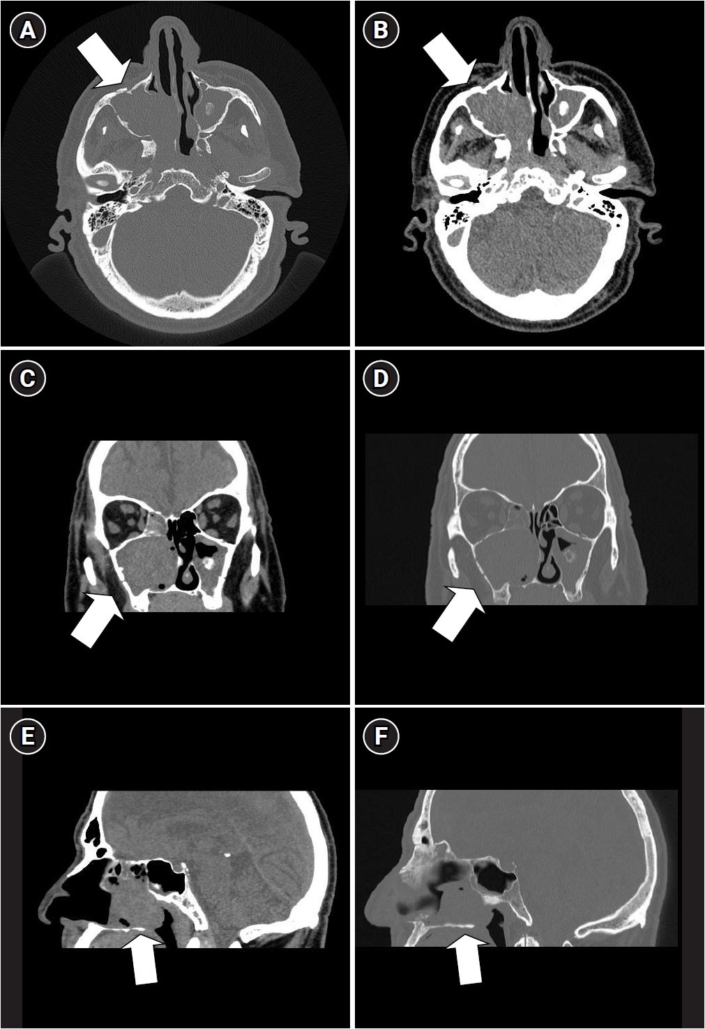
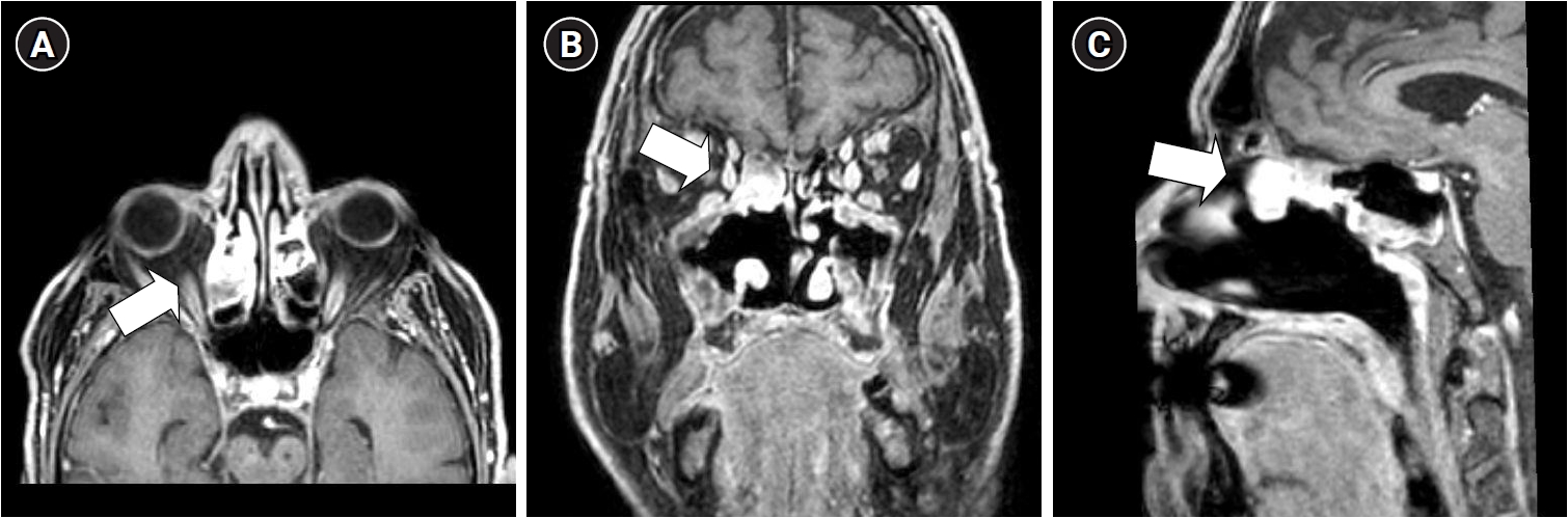
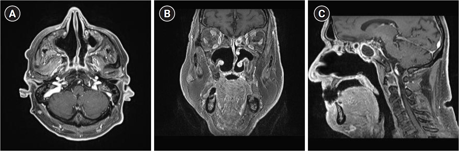
Fig. 1. Head computed tomography obtained on 13 Apr 2022. Axial (A, B), coronal (C, D), and sagittal (E, F) images showing an expansive lesion (about 5 cm across), occupying the right nasal-choanal cavity, the right ethmoidal cells and the right maxillary sinus, and eroding the medial sinus wall; evidence of an inflammatory process involving the left ethmoidal cells and the left maxillary sinus. The arrows point to the expansive lesion.
Fig. 2. Head magnetic resonance obtained on 10 Oct 2022. Contrast-enhanced axial (A), coronal (B), and sagittal (C) images (paramagnetic contrast medium), showing, on the right side, the persistence of a residual neoplastic lesion expanding towards the upper ethmoidal cells. The arrows point to the expansive lesion.
Fig. 3. Head magnetic resonance obtained on 1 Oct 2025. Contrast-enhanced axial (A), coronal (B), and sagittal (C) images (paramagnetic contrast medium), showing no persistence of residual neoplastic lesions.
Fig. 1.
Fig. 2.
Fig. 3.
Sinonasal hemangiosarcoma following juvenile occupational exposure to formaldehyde when breeding farm animals
| Calendar period | Age (years) | Relevant circumstance/event |
|---|---|---|
| 1967–1976 | 10–19 | Farmer and breeder on the family farm |
| Chemicals to which the subject was exposed: “verdigris” (a mixture of copper sulfate and calcium hydroxide), other fungicidal “sulfur products”, some not better identified insecticides, formaldehyde water-based solutions | ||
| 1977–2002 | 20–45 | “Carabiniere”, i.e., trooper of an Italian national military in charge of territorial control police duties |
| Chemicals to which the subject was exposed: none | ||
| 2019 | 62 | Onset of long-lasting nasal obstruction |
| 2022 March | 65 | Sudden emission of abundant, foul-smelling fluid |
| 2022 April | 65 | CT revealing a solid mass, about 5 cm across, occupying the right maxillary and ethmoidal sinuses |
| 2022 May | 65 | Surgery |
| 2022 Oct | 65 | MR recording the presence of residual cancer tissues along the margin of the surgical resection |
| 2022 Dec–2023 Feb | 65 | Proton therapy (33 sessions) |
| 2025 Oct | 68 | MR recording no residual cancer tissues |
| 2026 May | 69 | No evidence of a relapse of the disease |
Table 1. Occupational and disease timelines
CT: computed tomography; MR: magnetic resonance.


